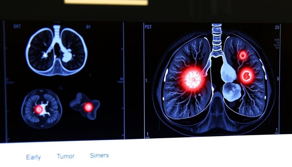
Lung cancer leads in causing deaths for both genders, more than breast, prostate, and colorectal cancers combined. This fact highlights the need for understanding lung cancer tumor images. They’re crucial for diagnosis and treatment plans. This guide sheds light on imaging techniques used in diagnosing lung cancer. It helps patients make better health decisions.
Key Takeaways
- Lung cancer remains the top cause of cancer-related deaths in the United States.
- 85% of lung cancer cases are diagnosed as non-small-cell lung cancer.
- Medical images are vital for early detection and appropriate treatment.
- Improved imaging techniques can lead to better prognosis outcomes.
- Patients benefit from understanding their imaging options, aiding informed conversations with healthcare providers.
Understanding Lung Cancer
Lung cancer significantly affects health, touching those diagnosed and their families. It is important to know what lung cancer is and its types. Most often, it starts with the uncontrollable growth of lung cells, which can form tumors. Knowing the different types of lung cancer is critical for choosing the right treatment.
Definition and Types of Lung Cancer
The main types of lung cancer are Non-Small Cell Lung Cancer (NSCLC) and Small Cell Lung Cancer (SCLC). NSCLC makes up about 85-90% of all cases. Its subtypes include adenocarcinoma, squamous cell carcinoma, and large cell carcinoma. SCLC, mostly caused by smoking, tends to spread fast. There are also carcinoid tumors and rare types like mesothelioma, linked to asbestos exposure.
Statistics and Mortality Rates
Lung cancer is the top cause of cancer deaths globally, leading to about 1.8 million deaths each year. The statistics show why it’s urgent to find the disease early and start treatment. Many cases are linked to smoking. But, it’s worth noting that about 15% of lung cancer cases are in non-smokers. This fact underscores the importance of research into other causes.
The Role of Medical Imaging in Lung Cancer Diagnosis
Medical imaging is key in spotting lung cancer early. This early detection is crucial for a better chance of successful treatment. Many imaging methods now help find lung cancer, all thanks to tech advancements.
The Importance of Early Detection
Finding lung cancer early can greatly improve the patient’s outlook. Using modern imaging tools helps catch the disease sooner. This leads to more effective treatments and a better life for those diagnosed early. Doctors urge people at risk to get scanned regularly.
Types of Imaging Techniques Used
Doctors use several types of scans to diagnose lung cancer:
- Chest X-rays: Often the first step, they can spot tumors but might miss small ones.
- Computed Tomography (CT) Scans: These scans use X-rays to make detailed pictures and are crucial for finding lung cancer.
- Magnetic Resonance Imaging (MRI): MRIs use radio waves and magnets to see if the cancer has spread to the brain or spinal cord.
- Positron Emission Tomography (PET) Scans: PET scans use a special dye to show where cancer is, helping doctors see if it has spread.
- PET/CT Scans: Combining PET and CT scans offers more detailed images for a more accurate diagnosis.
These imaging tools are essential in the fight against lung cancer. They help doctors get a full view of the patient’s condition. For the best lung cancer care, seeing a specialist is key. Click here for more info on top lung cancer doctors.
Types of Medical Images of Lung Cancer Tumors
Medical imaging is crucial when it comes to diagnosing lung cancer. It helps identify and stage tumors accurately. Different imaging techniques give essential information. This aids doctors in making the best decisions for patient care.
Chest X-rays: Initial Diagnosis
Chest X-rays are the first step in looking for lung cancer. They show a general picture and can spot obvious problems. This alerts doctors to the possibility of lung cancer. But, chest X-rays may mistake other issues for cancer or miss it. So, more detailed imaging is often needed for a reliable diagnosis.
CT Scans for Lung Nodules
CT scans give a much clearer picture than chest X-rays. These scans are key for checking and staging lung nodules. They let doctors see lung structures in detail. This helps in figuring out if nodules are cancerous or not and in planning treatment. Screening with low-dose CT scans has been effective in finding lung cancer early. Early detection leads to better chances of survival. Every year, about 1.35 million people get lung cancer worldwide.
MRI as a Secondary Approach
MRI is a second-level option for lung cancer diagnosis. It’s used in specific situations, like checking if a tumor has spread. But, MRI has issues, mainly with image clarity due to breathing. Yet, it can offer key insights for some treatment plans.
For deeper insight into imaging methods, visit this resource on radiological techniques about the lung. Well-rounded imaging is vital for identifying lung cancer types. It helps ensure patients get timely and proper treatment, improving their outcomes.
How CT Scans Aid in Lung Cancer Assessment
CT scan technology is very important in checking for lung cancer. It uses X-rays and computers to make detailed pictures of the lungs. These images help doctors spot small nodules, which might be lung cancer.
Understanding CT Scan Technology
CT scans are better than normal chest X-rays because they show more details. They tell doctors about a tumor’s size, shape, and place, and about nearby lymph nodes. This detailed information helps them figure out if a tumor is likely cancerous. If something looks off, a CT scan can lead to more tests. This means doctors can act swiftly and correctly.
Benefits of CT Scans in Early Detection
CT scans are very helpful in finding lung cancer early on. Studies show that using low-dose CT scans to screen for lung cancer lowers the death rate by 20% compared to old methods. These scans use a small amount of radiation but are still very useful for diagnosis. Finding cancer early improves chances of survival and helps in planning the best treatment. This leads to care that is more tailored to each patient.

Lung Tumor Segmentation Techniques
Lung tumor segmentation is key in lung cancer diagnosis and treatment planning. Medical imaging technology is advancing. Because of this, segmentation techniques for lung tumors are also getting better. Deep learning is changing the game in analyzing lung cancer images.
Conventional Segmentation Methods
Traditional segmentation involved manual work, which was slow and error-prone. Methods like thresholding, region growing, and edge detection were common. However, these methods had flaws due to how differently people might interpret the results.
This difference in interpretation could make diagnosing harder. It made planning treatment for patients more complicated.
Advancements in Deep Learning for Lung Cancer
Deep learning has changed how lung tumors are segmented. New deep learning tools, like U-Net and fully convolutional networks (FCNs), are very good at segmenting lung cancer images. They can spot and outline tumors accurately with less chance of mistakes.
One system showed a 97.83% accuracy in tumor segmentation. This was a big improvement over older methods. Transfer learning helps these systems work well, even with smaller data sets. This makes deep learning very useful in finding lung cancer.
Deep learning doesn’t just offer better accuracy but also speeds up image processing. This speed is vital for quick patient care. With these advanced models, early diagnosis becomes possible. This could stop lung cancer from getting worse.
AI-Assisted Lung Cancer Diagnosis
Artificial Intelligence is changing how we diagnose lung cancer. It uses something called convolutional neural networks. These networks dig deep into medical images to find lung cancer signs, faster and more reliably than older ways. AI help is crucial for radiologists. It makes sure they don’t miss important patterns and anomalies.
The Role of Convolutional Neural Networks
Convolutional neural networks (CNNs) are key for better lung cancer detection. These advanced models look through tons of medical image data. They identify important signs that point to cancer. AI tools have gotten a thumbs up from the Food and Drug Administration for finding various cancers, including lung cancer. These tools are better at telling non-cancerous growthes from cancerous ones.
Benefits of AI in Image Analysis
AI in image analysis brings several benefits to lung cancer diagnosis. It not only works faster and more precisely but also cuts down on wrong cancer diagnoses. These mistakes can cause a lot of worry for patients and cost more money. AI can scan quickly, taking minutes instead of days. It also helps doctors find urgent cases faster, leading to quicker treatment.

| Aspect | Traditional Methods | AI-Assisted Methods |
|---|---|---|
| Time for Diagnosis | Days | Minutes to Seconds |
| Accuracy in Detection | Varies | Up to 98.9% |
| Reduction in False Positives | High Rates | Significantly Lower |
| Utilization of Data | Limited | Extensive |
| Prioritization of Cases | Manual | Automated |
Importance of Lung Cancer Screening
Lung cancer screening is key for finding it early, especially in those at high risk. Screenings catch issues before they get worse. This can lead to better survival chances and results for at-risk folks.
Guidelines for Screening
Experts suggest screening mainly for older adults who smoked a lot over the years. They recommend yearly scans for people 55 to 80 years old who smoked heavily. This includes those who have quit smoking in the past 15 years.
To benefit most from screening, individuals should be healthy overall. That way, they can get the full advantages of detecting lung cancer early.
Risk Factors for Lung Cancer
Knowing what increases lung cancer risk is crucial for figuring out who should get screened. Here are some factors:
- Chronic obstructive pulmonary disease (COPD)
- A family history of lung cancer
- Exposure to asbestos or other carcinogens
- Long-term tobacco smoking
People with these risk factors are encouraged to get screened early. Early screening can make a big difference in treatment success. Recognizing these signs helps grasp the true value of lung cancer screenings.
Challenges in Imaging for Lung Cancer
Lung cancer imaging faces a lot of challenges that matter a lot for diagnosis and treatment. It’s vital to know these challenges. This knowledge helps make lung cancer imaging better and improves diagnosis.
Limitations of Current Imaging Techniques
Today’s imaging tools like CT and PET scans have big imaging limitations. Early-stage lung cancer is hard to spot accurately. Missing these early cases means some cancers are not found when they are small. Plus, telling different types of tumors apart is tough due to these limits. Nuclear medicine imaging also faces issues because of inconsistent results and other confusing factors.
The Need for Improved Imaging Technology
We need better improved lung cancer imaging technology. Using AI and deep learning could make radiographic analysis much sharper. AI helps turn qualitative assessments into precise, quantitative ones. This gives a clearer view of the tumor’s nature. Making imaging protocols uniform, especially in multicenter studies, is key for reliable results. It leads to better care for patients. EARL-compliant reconstruction is a way to make radiomics features more consistent, which could vastly improve lung cancer diagnosis accuracy.

| Imaging Type | Strengths | Limitations |
|---|---|---|
| CT Scans | High resolution, widely available | False negatives in small tumors |
| PET/CT | Effective for non-small cell lung cancer | Debate over small cell lung cancer accuracy |
| Nuclear Medicine Imaging | Useful in diagnosis and staging | Inconsistent results and specificity |
| AI-Enhanced Imaging | Increased accuracy in tumor characterization | Limited validation in clinical settings |
Research and Clinical Trials in Lung Cancer Imaging
The way we diagnose lung cancer is changing fast, thanks to research in lung cancer imaging and new clinical trials. Better imaging methods are being developed to find lung tumors more accurately. This is important for choosing the right treatment.
Current Innovations in Imaging
New imaging techniques focus on measuring tumors in more detail, moving past just looking at their size. Such techniques include volumetric CT and PET imaging. These are tested in clinical trials for lung cancer to learn more about the tumors. Over 5 million images from these studies are helping improve how we detect lung cancer. The FDA has also approved a new imaging drug called Cytalux (pafolacianine). It helps find cancers during surgery that other methods might miss.
Future Directions in Lung Cancer Diagnosis
The future looks bright for diagnosing lung cancer. Artificial intelligence may soon tailor imaging to fit each patient’s needs better, making diagnoses more accurate. In the ELUCIDATE trial, advanced imaging spotted cancers in half of the patients that were previously missed. The goal of ongoing research in lung cancer imaging is to make these technologies even better, leading to better care for patients.
Conclusion
Studying medical images of lung cancer shows how vital early detection is. It can greatly improve patient outcomes. Although the overall 5-year survival rate for lung cancer is around 15%, there’s hope. For T1a tumors, the survival rate can reach 77% with early detection.
Recent advances in imaging tech and AI have brought new hope in diagnosing lung cancer. Deep learning models, for example, have shown high accuracy in spotting lung cancer. These advances support better treatment plans, helping patients greatly.
It’s key for healthcare pros and patients to keep up with lung cancer screening info. If considering screening, it’s wise to review the US Preventive Services Task Force guidelines. This article’s insights reinforce the need for early detection and new methods in combating lung cancer.