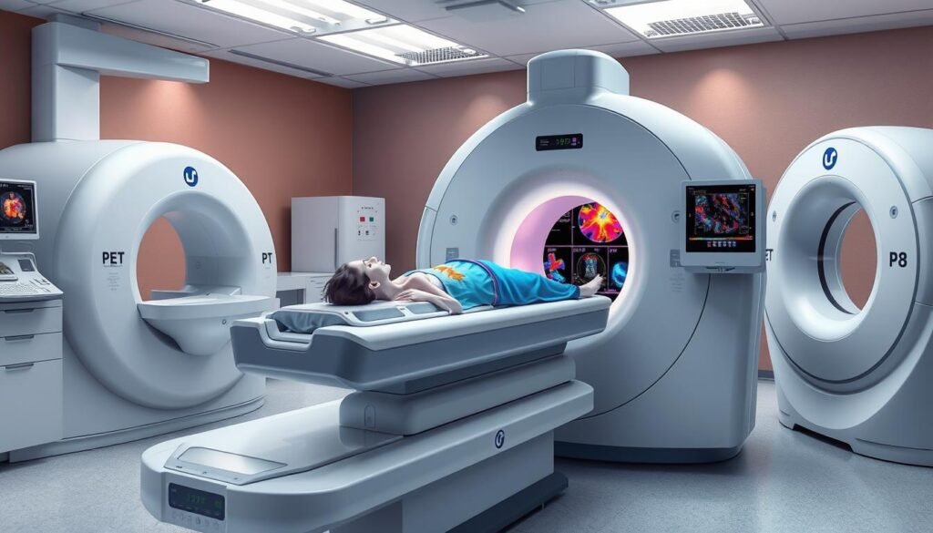
PET scans play a big role in finding and assessing many health issues. They last 20 to 40 minutes. These scans are really important for spotting cancer early on. They help doctors see if tumors are there and how far the disease has spread.
This piece will make PET scan results easier to understand. It will talk about what the results mean and why they are important. Knowing how PET scans are read can help you talk better with your doctor. This can lead to making smarter choices about your health. Getting what the results show about cell activity and how the disease is staged is key.
Key Takeaways
- PET scans are crucial for cancer diagnosis and disease staging.
- The most commonly used radiotracer in PET scans is fluorodeoxyglucose (FDG).
- Understanding results can require discussions about metabolic activity and SUV levels.
- Insights from PET scan analysis can guide treatment options in various medical disciplines.
- Combining PET scans with CT or MRI can enhance diagnostic accuracy.
What is a PET Scan?
A positron emission tomography (PET) scan offers a deep look into our body’s workings. It marks a big step forward in medical imaging. Unlike CT or MRI scans that show us the structure, PET scans reveal how our tissues work. They do this by tracking how tissues take in a special radioactive tracer. This shines a light on uncommon metabolic activities that could point to health problems.
Here’s how it works. A tiny amount of radioactive tracer goes into your vein. Your organs and tissues soak it up over about an hour. This lets the scan show us areas with a lot of metabolic activity. Such detailed images help in finding cancers, checking heart health, and understanding brain issues.
Sometimes, doctors combine PET with CT scans for something called a PET/CT scan. This helps them locate tumors more accurately. The radiation in a PET scan is about the same as in most CT scans. Normally, your body gets rid of the radioactive material in 2 to 10 hours.
PET scans play a key role in identifying and keeping track of many health conditions. However, sometimes they give false results. Blood sugar levels or insulin use in those with diabetes can affect the outcomes. Though rare, some people may have allergic reactions to the tracer. This could show up as pain, redness, or swelling where they got the shot.
Want to know more about how PET scans help in diagnosing diseases? Check out this resource.
How Does a PET Scan Work?
A PET scan begins with a person resting on a table that moves into a big machine. Before the scan, they receive a radioactive tracer through an arm vein. This tracer travels in the body, gathering in active areas.
After waiting for the tracer to spread, the machine takes images. These images highlight the body’s metabolic activity in different organs and parts.
PET scans show detailed metabolic activity by tracing glucose. This can point to diseases like cancer. They can show cell changes that MRIs and CT scans might miss. This is crucial for diagnosing complex diseases.
In the U.S., doctors perform over 2 million PET scans every year. They’re essential for finding cancer and assessing brain and heart diseases. For instance, a tracer called fluorodeoxyglucose (FDG) reveals significant metabolic hotspots.
The PET scan is safe, following strict FDA radiation rules. However, pregnant folks should avoid it to protect their unborn babies. Doctors also check for allergies or conditions like kidney disease that could affect the scan. For radiation therapy information, you can read this guide.
The Role of Radiotracers in PET Scans
Radiotracers play a key role in PET scans by highlighting areas with unusual metabolic rates. They use radioactive isotopes to show various biological processes. A top tracer in clinics is Fluorodeoxyglucose (FDG).
FDG acts like glucose but is drawn to cells using lots of glucose, often seen in cancer. This feature makes FDG vital in spotting cancers in the body.
FDG can map out how cancer spreads in many types, like cervical and lung cancer. It shows us how cancer cells change to use more glucose. This is called the Warburg effect.
Yet, FDG isn’t just for spotting cancer. Some non-cancerous activities can also pick it up. This can happen due to inflammation or natural organ activities. This sometimes makes reading the results tough. But FDG PET is still key in cancer care.
Beyond FDG, new radiotracers are being looked into. For example, 18F-sodium fluoride (18F-NaF) helps with checking bone metabolism. There are also tests on FAPI PET imaging agents for various cancers.
Knowing how different tracers work in PET scans is crucial for both doctors and patients. While FDG is a major tool, new techniques are broadening our view. They help improve how we diagnose diseases. Below is a chart comparing PET tracers and what they do:
| Radiotracer | Application | Common Cancers | Note |
|---|---|---|---|
| FDG | Metabolic activity | Cervical, Colorectal, Esophageal, Lung | Nonspecific uptake in inflammation possible |
| 18F-NaF | Bone metabolism | Bone metastases | FDA-approved for clinical use |
| [68Ga]Ga-FAPI-04 | Tumor detection | Various cancers | High tumor to background ratios observed |
| [18F]FLT | Tumor aggressiveness | Ovarian, Lymphoma | Better staging correlation in soft tissue tumors |
Understanding Different Imaging Techniques
In the field of cancer diagnosis, various imaging techniques are vital. Positron Emission Tomography (PET) is often chosen to look at metabolic activity, especially in cancer cases. It lets doctors see biochemical processes in tissues, which is key for finding cancer.
Then there’s Computed Tomography (CT), which uses X-rays for detailed images of the body inside. CT scans give us a clear view of tumors’ size, shape, and place. On the other side, Magnetic Resonance Imaging (MRI) uses magnetic fields and radio waves. It gives detailed images of soft tissues, helping see tumor margins and nearby areas.
When PET and CT are combined into PET-CT, the results are even more precise. This mix of metabolic and anatomical imaging is crucial in cancer care. It helps with:
- Initial cancer staging
- Assessing the spread of cancer
- Evaluating treatment efficacy
PET-CT and PET-MRI tests both use 18F-fluorodeoxyglucose (FDG). This helps find metabolism changes related to cancer. Combining metabolic with anatomical imaging gives a full picture of a patient’s condition.
Preparing patients for these imaging techniques is key for good results. Not doing physical activities before the test and fasting can lead to better imaging. This lowers the chance of unclear results.
| Imaging Modality | Primary Use | Strengths | Limitations |
|---|---|---|---|
| PET | Metabolic activity assessment | High sensitivity to biochemical changes | Lower spatial resolution |
| CT | Structural imaging | Detailed anatomical views | Limited functional information |
| MRI | Soft tissue imaging | Excellent contrast for soft tissues | Longer acquisition times, higher costs |

Interpreting PET Scan Results
Understanding PET scan results can be tough due to medical jargon. Many people find the report’s details hard, especially the complex terms. Learning some common terms helps talk better with doctors and understand the findings. This talk is key to knowing what the results mean.
Common Terms in PET Scan Reports
In PET scan reports, some terms are very common. Each term has a specific meaning that’s important for understanding the report:
- Uptake Values: Show how much radiotracer tissue takes in; high levels may mean more metabolic activity.
- Standard Uptake Value (SUV): A measure for comparing radiotracer uptake, making it easier to spot issues.
- Physiologic Uptake: This is the normal activity in tissues, used as a comparison point.
Knowing these terms means you can ask better questions when talking to your doctor. This makes the whole process less confusing.
Engaging with Your Doctor about Results
After getting a PET scan, talking to your doctor is key. There are a few reasons why it’s so important:
- It clears up any confusion about the report.
- It outlines what comes next, whether it’s more tests or treatments.
- It lets patients play a part in their care, making choices with more knowledge.
Doctors go through the scans in detail with patients. This helps you understand what the findings mean. It also tells you what might happen next, based on those results.
Metabolic Activity Indicated by FDG Uptake
Understanding how FDG uptake shows metabolic activity is key in spotting cancer. FDG helps us see how cells use glucose. This can tell us if there’s unusual activity, like cancer, or just normal, healthy function. Seeing early abnormal FDG uptake on PET scans could point to cancer or other metabolic issues.
What is FDG and Its Significance?
FDG is the top PET tracer used in finding cancer, approved by the FDA. It’s behind over 90% of PET scans in oncology because it highlights increased metabolic activity, often seen in cancer cells. Cancer cells often eat up glucose faster, even with enough oxygen around. This is why FDG is so valuable in detecting cancer.
How to Determine Normal FDG Uptake Levels
The normal rate of FDG uptake differs across tissues. The brain and liver usually show more activity because they need lots of energy. It’s important to know what’s normal to spot anything suspicious. Things like age and blood sugar levels can also impact FDG uptake, so doctors keep an eye on these.
| Tissue Type | Normal FDG Uptake Range |
|---|---|
| Brain | High |
| Liver | Moderate to High |
| Skeletal Muscle | Low to Moderate |
| Heart | Moderate |
| Kidney | Variable |
Understanding FDG uptake levels accurately is critical for the right read of PET scan outcomes. For more details on FDG uptake, check out this link here.

Evaluating Tumor Detection through PET Scans
PET imaging has become key in spotting tumors in cancer care. It started almost 30 years ago and has changed how we see cancer. Positron emission tomography shows where cells are more active, pointing to possible cancer. This helps doctors tell non-cancerous growths from cancerous ones, improving treatment.
Today, many PET scanners are used worldwide. Medicare sees their value in identifying various cancers. These include lung, colorectal, melanoma, and lymphoma. But, PET scans don’t work as well for some cancers, like those in the breast and thyroid.
Tumors react differently to PET scans because each type uses glucose differently. Some tumors with low glucose activity might not show up well on a PET scan. This shows why we need better ways to look at cancer.
Modern PET scanners have up to 24 rings of detectors. This allows them to take detailed images. With these images, doctors can better predict how a patient will respond to treatment. They often get it right about 70% of the time.
Special techniques help match initial scans with later ones for tracking tumor changes. This helps doctors see how the tumor responds over time. Tools like PERCIST and the SUV metric help in planning treatment.
PET imaging is crucial not just for finding tumors but also for checking if cancer comes back. It remains vital in caring for cancer patients. With ongoing advancements, PET scans are set to offer even more help to those fighting cancer.
| Cancer Type | FDG-PET Indication | Considerations |
|---|---|---|
| Non-Small Cell Lung Cancer | Approved | Standard metabolism rates |
| Colorectal Cancer | Approved | High glucose uptake |
| Melanoma | Approved | Variable uptake patterns |
| Lymphoma | Approved | Effective for staging |
| Breast Cancer | Restricted | Low sensitivity and specificity |
| Thyroid Cancer | Restricted | Alternative imaging recommended |
Clinical Applications of PET Scans in Oncology
PET scans are crucial in today’s cancer care. They do more than just find cancer. These scans use a radioactive tracer to see how organs work, spotting cell changes earlier than other scans. PET-CT scans offer 3D views for better diagnosis.
Doctors use PET scans to spot cancer, check if treatment works, and predict outcomes. Oncologists rely on them for real-time tumor activity, adjusting treatment as needed. This makes cancer care more personalized and effective.
But PET scans aren’t just for cancer. They also check for brain issues like dementia and epilepsy. Advanced PET/MRI tech is great for diagnosing soft tissue cancer. So, PET imaging’s versatility is key in oncology.
PET scans are mostly safe, though they have risks due to the radioactive tracers. This is a concern for pregnant or nursing women and diabetics. Rology’s teleradiology makes reading PET scans easier, providing expert advice anytime.
From May 2020 to June 2023, many PET/CT exams highlighted their role in oncology. They mainly found nasopharyngeal carcinoma, lymphoma, and lung cancer in adults aged 45-65. Children with cancer like lymphoma and neuroblastoma get special care.
PET imaging’s wide use in cancer treatment shows how vital it is for patient care.
Read more about PET scans in oncology here.
Understanding Disease Staging with PET Imaging
PET imaging is key to staging diseases, especially cancer. It helps by showing how far cancer has spread. This lets doctors personalize treatment plans.
FDG PET scans are great at checking tumors. They focus on how cells use energy, spotting cancer cells that might not show up on other scans. This is because cancer cells use more energy, lighting up brightly on scans.
It’s important to understand FDG uptake details. The uptake value shows the tumor’s radiotracer absorption, revealing its aggression and metabolic rate. Nuclear medicine experts often review these findings for better clarity.
PET imaging also helps in tracking how well treatment works. Seeing changes in the tumor early on helps in improving care. This leads to better results for patients.
In conclusion, PET imaging is crucial for detailed disease staging. It shows cell activity that other methods might miss, making it vital in fighting cancer.
| Category | Description |
|---|---|
| Purpose | Assess the extent of cancer spread and guide treatment decisions. |
| Key Radiotracer | [F-18] fluorodeoxyglucose (FDG) is the most commonly used. |
| Metabolic Activity | Cancer cells show higher metabolic rates, appearing as bright spots on scans. |
| Metrics | Standardized uptake value (SUV) used to evaluate tumor behavior over time. |
| Second Opinions | Highly recommended for accurate interpretation of complex results. |
| Detection Limits | Up to 20% of tumors less than 15mm may go undetected. |
Recent Advancements in PET Scan Analysis
PET imaging technology has made huge strides, especially in finding cancer. Now, we have better imaging algorithms and new radiotracers. These advancements make PET scans more precise, helping doctors find and describe diseases earlier and more accurately.
New technologies, like PET-MRI, are changing how we diagnose and monitor cancer. PET-MRI combines metabolic and structural details to give a fuller picture. Also, dual-energy CT makes PET images more quantitatively accurate. These improvements are upgrading treatment plans too.

There’s also progress in creating quicker, cheaper scintillators for better imaging. Solid-state photodetectors are now cheaper and can work with MRI. These developments are making PET scans more accessible and useful.
Today, research includes multi-isotope PET imaging for tracking different bodily processes at once. 4D imaging is expanding into areas like cardiology for both preclinical and some clinical use. These steps forward are helping doctors understand cancers better and tailor treatments to patients.
If you’re interested in better ways to catch cancer early, combining imaging with new blood tests is promising. Find out more by visiting this resource for the latest research.
Conclusion
It’s very important for patients to understand PET scans when they are dealing with cancer. Learning about how PET scans work and the role of radiotracers like FDG helps. It also helps to know how to read the results. This info makes talking with doctors easier and helps patients make good choices about their health.
New advancements, like PSMA ligands Gallium PSMA 11 and FDA-approved DCFPyL, show how vital PET scans are. The use of PSMA-RADS to measure prostate cancer risk shows the value of these scans. Plus, having standard ways to report results helps improve patient care.
PET scans have become a key part in fighting cancer. They help in finding out how bad the cancer is and the best way to treat it. If patients understand all this, they can help make decisions about their treatment. This team effort leads to better health results.