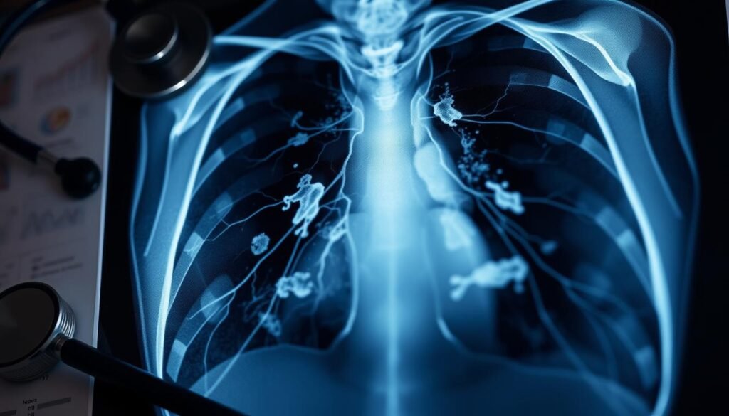
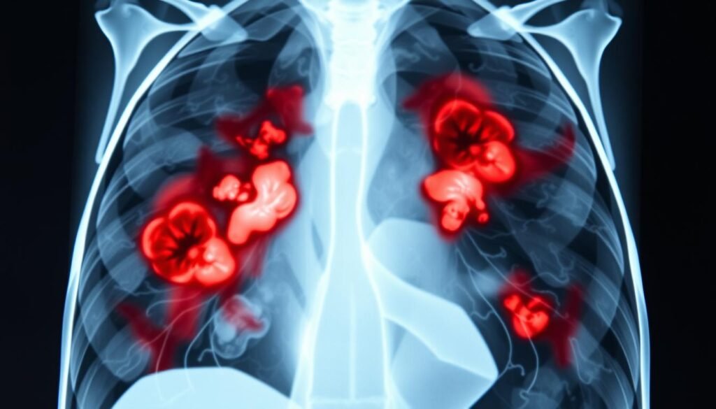
Lung cancer leads in causing cancer-related deaths across men and women in the U.S. This fact stresses the need for detecting lung cancer early, boosting the chances of survival. Through lung cancer X rays, doctors can better identify and understand the disease, leading to better care for patients.
This guide offers a detailed look at lung cancer X rays. It explains how they help in diagnosing the disease and affects patient treatment. We also look into modern diagnosis methods and why they’re crucial. For more details, visit this resource.
Key Takeaways
- Lung cancer remains the top cause of cancer deaths in the U.S.
- Utilizing lung cancer X rays aids in early lung cancer diagnosis.
- Early detection is crucial for improving treatment outcomes.
- Common risk factors include smoking and exposure to harmful substances.
- Understanding the different types of lung cancer is vital for effective management.
Introduction to Lung Cancer
Lung cancer is the second most common cancer in the United States. It causes the most cancer deaths each year. This fact highlights the importance of lung cancer awareness. Smoking is the main cause of lung cancer. Roughly 80-90% of those with lung cancer have smoked at some point.
Knowing the risks of lung cancer is vital for stopping it early. If you are between 50 and 80 years old and have a history of smoking, get a yearly low-dose CT scan. This scan saves lives by catching lung cancer early in people at high risk. However, it’s not perfect and might not catch every case. It could also lead to extra tests that are not needed.
Every year, lung cancer affects 1.35 million people worldwide and causes 1.8 million deaths. The most common kind, non-small cell lung cancer (NSCLC), has different types like squamous cell carcinoma. This type is about 30% of cases. It’s key to know the symptoms and risks linked to smoking and lung cancer. We can fight this deadly disease through better public health actions and more awareness.
Understanding Lung Cancer X Ray
A lung cancer X-ray is key for looking at lung parts. It is first used when checking for lung cancer. Besides showing tumors, it can reveal things like lesions and fluid build-up.
What is a Lung Cancer X Ray?
A chest X-ray shows different shades depending on the tissue. Tumors in the lung look like white-gray areas. But, it’s not perfect for diagnosing lung cancer by itself. It sometimes can’t tell cancer from other lung problems, needing more tests.
How Lung Cancer X Ray is Used in Diagnosis
Chest X-ray results often lead to more tests. CT scans come next, providing clearer images. If CT scans suggest cancer, a PET-CT scan is done for detailed info. This helps make good treatment choices.
X-rays help spot possible lung nodules, but can’t confirm cancer alone. With newer, larger imaging tools, chest X-rays aren’t the only option anymore.
If you’re worried about lung cancer, understanding X-rays is a good step. Knowing their limits and the need for more testing can guide lung health choices. To learn more, check out this source.
Risk Factors Associated with Lung Cancer
Learning about lung cancer risk factors helps with prevention. Some factors raise the risk of this disease. Knowing about personal and environmental impacts encourages healthier living.
The Role of Smoking
Smoking is the top cause of lung cancer, behind about 90% of cases. It’s riskier for those who smoke more and longer. Even non-smokers can get lung cancer, showing how critical smoking is in lung cancer risk.
Hardcore smokers mostly get small cell lung cancer. Other types, like squamous cell and adenocarcinoma, fall under non-small cell lung cancer.
Other Environmental Risks
Besides smoking, other things like secondhand smoke and radon gas add to the risk. Jobs in mining or construction can be dangerous too. Air pollution and indoor carcinogens are harmful as well. Reducing these risks is a smart move. For more, visit this source.
Genetic and Family History Factors
Genetics also play a role in lung cancer risk. Having lung cancer in the family may increase your risk. It’s essential to understand these factors for prevention and awareness.
The Importance of Early Lung Cancer Detection
Early detection of lung cancer is key to better treatment results and longer lives. Spotting symptoms early leads to timely help, which greatly improves a patient’s future. Studies show that detecting lung cancer early greatly raises the chances of successful treatment.
How Early Detection Improves Outcomes
For those at high risk, tests like low-dose computed tomography (LDCT) are suggested. Adults 50 to 80 with a history of smoking should get checked every year, says the US Preventive Services Task Force. This leads to fewer deaths from lung cancer and healthier lungs. People who get LDCT tests have a better chance of surviving than those who find out later.
Signs and Symptoms of Early Lung Cancer
Know the early signs of lung cancer for better detection. Look out for:
- Persistent cough
- Chest pain
- Weight loss without explanation
- Coughing up blood
- Shortness of breath
Catching these signs early can mean faster treatment and better chances for those with lung cancer. Teaching people about these signs and the importance of early detection saves lives.

Chest X-Ray Imaging Techniques
Chest x-ray imaging is key in assessing lung cancer. It offers valuable insights into lung health. The core methods include both frontal and lateral views. These provide a detailed look at lung conditions and structures.
Standard Techniques for Lung Assessment
Chest x-rays are vital for spotting lung issues, used widely in healthcare. They help identify lung infections, pneumonia, tuberculosis, lung cancer, enlarged heart, and blocked blood vessels.
- Lung infections
- Pneumonia
- Tuberculosis
- Lung cancer
- Enlarged heart
- Blocked blood vessels
Digital systems improve image quality and may lower radiation risks. Images are ready quickly and reviewed by a specialist.
Limitations of Chest X-Rays in Lung Cancer Diagnosis
While crucial, chest x-rays have limitations. They struggle to detect early-stage cancer and to tell non-cancerous from cancerous nodules. They also can’t show if the cancer has spread to nearby areas.
Studies show they don’t much reduce lung cancer deaths. CT scans, on the other hand, offer better images and can detect early lung cancer.
| Parameter | Chest X-Ray | Computed Tomography (CT) |
|---|---|---|
| Image Quality | Standard | High |
| Detection of Early Lung Cancer | Limited | Enhanced |
| Radiation Exposure | Low | Higher |
| Diagnosis of Tumor Invasion | Not Possible | Possible with Advanced Techniques |
Knowing these differences helps doctors choose the best method for examining lung cancer. This ensures a full review of a patient’s health.
Radiographic Screening for Lung Cancer
Different methods for spotting lung cancer early are key. Chest X-rays and low-dose CT scans are often compared. This is because they differ in how well and safely they work.
Comparison of Chest X-Ray and CT Scans
Chest X-rays were the go-to for spotting lung issues. But studies show low-dose CT scans catch early lung cancer better. A big study found people scanned with LDCT had a higher chance of living longer than those checked with X-rays.
CT scans are better at finding small growths with less radiation. A low-dose CT scan uses much less radiation, making it safer for regular checks. Still, there is a downside. They can give false alarms, causing worry and more tests.
| Screening Method | Detection Capability | Radiation Exposure | False Positive Rate |
|---|---|---|---|
| Chest X-Ray | Lower | Higher | Moderate |
| Low-Dose CT Scan | Higher | Lower | Higher |
Recommendations for High-Risk Populations
The U.S. Preventive Services Task Force suggests yearly checks with LDCT for those 50 to 80. This is for people who smoked a lot or quit in the last 15 years. Many hospitals in the U.S. have set up screening programs for these high-risk groups.
This method helps catch lung cancer early, possibly lowering deaths among those at risk. Doctors need to highlight the screening’s benefits but also talk about its downsides. These include false alarms that can lead to worry.

Interpreting Lung Cancer X Ray Results
Understanding lung cancer X-ray results is key in managing lung health. Radiologists look for signs of possible cancer. These signs help decide if more tests are needed.
Common Indicators of Lung Cancer
Several X-ray signs may hint at lung cancer. These signs include:
- Spiculated masses: Irregular mass edges may mean a higher chance of cancer.
- Abnormal shadows: These might show possible tumors, which need more checks.
- Enlarged lymph nodes: This could mean cancer has spread from the lungs.
The first chest X-ray is an early step to find lung issues. But, CT scans give a clearer view of lung tumors.
Identifying Pulmonary Nodules on X-Rays
Finding pulmonary nodules is crucial in reading lung X-rays. A nodule is a small lung growth, which can be benign or malignant. Examples include:
| Example | Description |
|---|---|
| New Indeterminate Nodule | A new nodule in the left lung, requiring a chest CT. |
| Pneumonia | Pneumonia in the right lower lobe, with a tip to check again in 4-6 weeks. |
Spotting these nodules and knowing their features impacts patient care. Detailed imaging results are crucial for precise lung X-ray interpretations.
Understanding Different Types of Lung Cancer
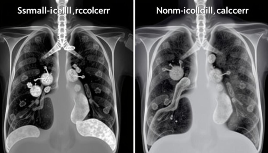
Lung cancers are mainly split into two types: small-cell lung cancer and non-small cell lung cancer. Knowing the differences is key for treatment and improving patient chances.
Small-Cell Lung Cancer vs. Non-Small Cell Lung Cancer
Small-cell lung cancer (SCLC) is known for being very aggressive and is often caused by cigarette smoking. On the other hand, non-small cell lung cancer (NSCLC) is more common, making up about 85% of cases. NSCLC grows more slowly than SCLC and has three main varieties:
- Adenocarcinoma: This type is most common in the U.S., especially among non-smokers. It makes up 40% of NSCLC cases and is found in the lungs’ outer areas.
- Squamous Cell Carcinoma: Found near the airways in the central lungs, this subtype makes up about 30% of NSCLC cases.
- Large Cell Carcinoma: This less common form grows and spreads faster than adenocarcinoma or squamous cell carcinoma.
Characteristics of Various Lung Cancer Types
There are many types of lung cancer, each with unique characteristics. Carcinoid tumors are one example, being either typical or atypical, with the typical kind being 90% of such cases. Another type, mesothelioma, makes up about 5% of lung cancer cases and is associated with asbestos exposure.
It’s crucial to understand each cancer type for better treatment approaches. Treatments can include surgery, chemotherapy, and radiation, all tailored to the cancer’s specific type and stage. Regular biomarker testing is also vital. It helps find certain mutations or proteins, leading to more personalized treatment plans and better outcomes for patients.

Follow-Up Procedures After a Suspected Diagnosis
After a lung cancer suspicion, follow-up steps are key for confirming the diagnosis and shaping treatment. These include various diagnostic tests for lung cancer, like biopsies. Understanding these tests helps patients and doctors make better decisions.
Biopsies and Other Diagnostic Tests
Biopsies are vital in evaluating lung cancer. The chance of lung nodules being cancerous depends on many factors. These include the nodule’s size, tissue density, and location in the lungs. Also, the nodule’s growth and the patient’s history matter.
If the results raise concerns, doctors may suggest earlier follow-up scans. The Lung CT Screening Reporting & Data System® (Lung-RADS®) by the American College of Radiology helps manage these diagnostics.
Role of Thoracic Radiology in Lung Cancer Management
Thoracic radiology is crucial in lung cancer care. It helps in staging the cancer, which is key for planning treatment. Through advanced imaging, radiologists can pinpoint which nodules may need more checks and help plan biopsies.
However, not everyone follows up as needed after a positive screening. Only about 42.6% get the follow-up care required. This shows how vital clear communication and understanding the need for prompt follow-up are.
| Lung-RADS Category | Adherence Rate (%) |
|---|---|
| Lung-RADS 3 | 30.0 |
| Lung-RADS 4A | 49.5 |
| Lung-RADS 4B or 4X | 68.0 |
Knowning how to follow up after lung cancer suspicion is crucial. It helps patients and doctors work together effectively. This step is key for the best outcomes and successful treatments.
For more info, check this lung cancer guide.
Conclusion
Lung cancer awareness is key to bettering patient outcomes. It helps find the disease early and treat it effectively. Chest X-rays are crucial for diagnosis and are often used in check-ups. A study found that 66% of lung cancer patients had an X-ray before their diagnosis in that year.
This fact shows why regular screenings are critical. Even with 23% showing no signs on X-rays, early detection chances grow closer to the diagnosis date. But, symptoms may not always be noticeable on X-rays. In 10% of cases, X-rays looked normal.
This means we need better ways to find lung cancer early. The accuracy of X-rays in showing symptoms is around 76.8% to 79.7%. By knowing more about what causes lung cancer and its signs, doctors can catch it sooner. This leads to better chances of surviving.
Ending on a note of action, spreading the word on lung cancer and encouraging regular checks is vital in fighting this illness. The survival rate has been improving slowly over the years, now up to 10% since 1971. Yet, improving how we fight lung cancer remains crucial. We must keep teaching people and reaching out to the community. This will help catch the disease early and improve life for those with lung cancer.