Did you know that about 70% of lung cancer cases are found after symptoms start showing? This makes finding it early tough. Small cell lung cancer (SCLC) makes up around 13-15% of all lung cancer types. Its fast growth can drastically shorten life if not treated quickly. Early lung cancer screening is vital for catching SCLC.
Chest X-rays are often the first step in spotting SCLC. They help doctors see unusual spots in the lungs early on. This can lead to faster treatment and better chances for patients. Knowing about SCLC and how doctors use imaging to find it is key for the best care.
Key Takeaways
- Small cell lung cancer represents a significant challenge in early detection.
- Most lung cancers are found through symptoms rather than routine screening.
- Chest radiography often serves as the initial lung examination tool.
- Advanced imaging techniques like CT and MRI improve diagnostic accuracy.
- Timely intervention can significantly enhance treatment outcomes.
- For more detailed insights, refer to sources on lung cancer diagnosis and management here.
Understanding Small Cell Lung Cancer (SCLC)
Small Cell Lung Cancer (SCLC) accounts for about 15% of all lung cancer cases. It’s known as a major type of lung cancer. The disease comes in two main stages: limited and extensive.
In the limited stage, cancer stays in one lung and may reach nearby lymph nodes. When it’s called extensive stage, it means the cancer spread to the other lung or even farther. This includes distant lymph nodes or other organs.
People who smoke are more likely to get SCLC, making it rare in non-smokers. Sadly, survival rates are not very high. On average, patients live about 12 months after being diagnosed. It’s crucial to understand SCLC well to plan treatments effectively. This is because surgery isn’t usually an option due to the cancer’s rapid spread at the time of diagnosis.
Treatment mainly involves chemotherapy, radiation, and immunotherapy. Chemotherapy is a key treatment, given in cycles. Radiation therapy is often used with chemotherapy in limited-stage SCLC. It targets the tumor and lymph nodes. Immunotherapy helps boost the immune system to fight the cancer cells.
Despite the difficulties SCLC presents, new treatments are being researched. Ongoing studies and clinical trials are crucial. They help doctors find better ways to treat this cancer. Healthcare professionals need a deep understanding of SCLC to improve patient outcomes significantly.
Importance of Early Cancer Detection
Finding cancer early is key to increasing survival chances for lung cancer patients. Lung cancer is the number two most common cancer and early action can change results majorly. People aged 50 to 80 who have smoked a lot are in the high-risk group. They are ideal for low-dose CT (LDCT) scans, better than usual chest X-rays.
Research shows LDCT scans find lung cancer early, which lowers death rates. While usual chest X-rays don’t help much in finding lung cancer or extending life, LDCT scanning does. Finding cancer early is vital because it means quicker treatment.
However, those getting scanned should know LDCT scans aren’t perfect. They might miss some lung cancers and show things that aren’t cancer. This could mean more tests and more radiation. Having modern CT tech and skilled specialists at screening centers is crucial for the best care and follow-up.
Good lung cancer screening helps catch the disease early. It also highlights how important it is to stop smoking since quitting greatly lowers your lung cancer risk. Thankfully, most health insurance plans, like Medicare, pay for lung cancer screening. This shows it’s a big part of stopping cancer before it starts.
Chest Radiography and Its Role in Detection
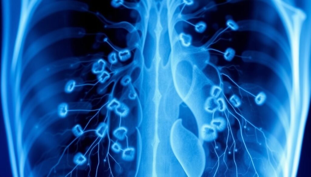
Chest radiography, or chest X-ray, is the first test done to check lung issues. It’s a simple way to see the lungs, heart, and chest bones. It’s good at finding big tumors and other lung problems. However, it’s crucial to know its limits for correctly diagnosing lung cancer.
How Chest X-rays Work
When getting a chest X-ray, doctors get a 2D picture of your chest area. This black and white image shows the inside organs. It helps find lung problems based on their size. But, it’s tough to spot smaller cancers, under 1.5 cm. Research says many lung cancers, like adenocarcinoma, might not be seen because of where they are or because other body parts hide them.
Limitations of Chest Radiography
Even though chest X-rays are common, they’re not perfect at finding lung cancer. One big problem is they can miss cancer in people without symptoms. Studies have found that 20% to 23% of X-rays can wrongly show no cancer in patients with lung cancer signs. Thus, many cancers aren’t found until they’re far along. That’s why doctors often suggest CT scans, especially for those at greater risk.
Imaging Techniques for Small Cell Lung Cancer
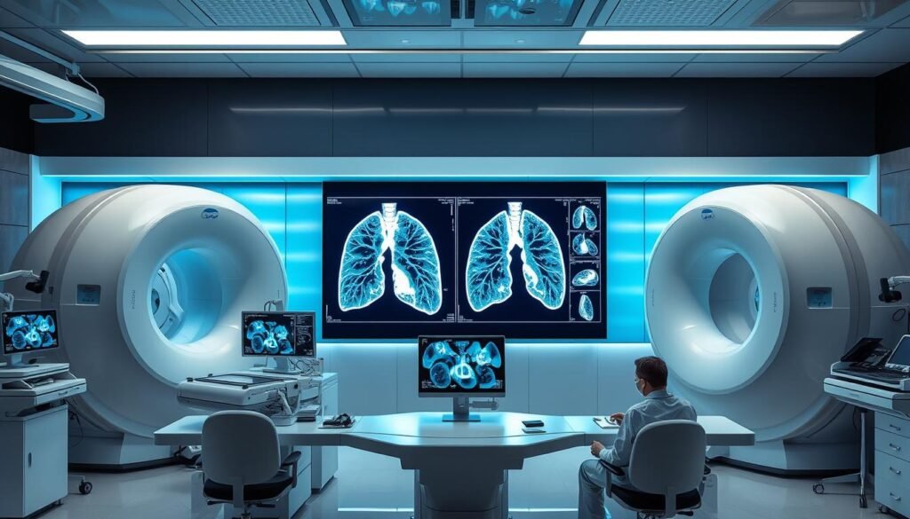
Advanced imaging techniques are key in managing small cell lung cancer (SCLC). Each method gives unique insights for better diagnosis and treatment. We’ll look at three main imaging techniques: CT scans, MRI scans, and PET scans.
Computed Tomography (CT) Scans
CT scans are top choice for checking lung cancer. They show clear images of the lungs. This helps identify lung masses and possible spread.
The use of multi-detector computed tomography (MDCT) stands out. Its speed and detail are great for spotting and studying tumors. Using contrast with CT scans provides vital info. This helps accurately stage SCLC.
Magnetic Resonance Imaging (MRI) Scans
MRI scans are crucial for seeing if small cell lung cancer has spread. They are particularly helpful for spotting potential brain metastases. About 20 to 30 percent of patients show brain involvement at diagnosis.
MRI offers detailed images of soft tissues. This boosts our understanding of how the disease evolves.
Positron Emission Tomography (PET) Scans
PET scans are very helpful for staging and planning treatment for SCLC patients. They check the cancer’s metabolic activity. This can show active disease areas that other scans might miss.
When PET and CT scans are used together, the accuracy of staging improves. This ensures a full view of the cancer. It also guides treatment plans well.
Small Cell Lung Cancer Xray: Detection and Diagnosis
Early detection is key in treating small cell lung cancer (SCLC). Knowing the radiographic characteristics on an X-ray is important. This knowledge can greatly affect the treatment results. Chest X-rays are usually the first step in diagnosing, showing signs that might point to this fast-moving cancer.
Common Findings on Chest X-rays
In chest X-rays, small cell lung cancer often shows as a lung mass in the center of the chest. It may come with:
- Pleural effusions
- Mediastinal lymphadenopathy
- Occluded bronchi caused by large masses
These signs can lead to more scans and tests. Regular check-ups can spot early signs like a lasting cough or unexpected weight loss. These symptoms could make your doctor consider a chest X-ray. You can learn about early signs in this useful link.
Radiographic Characteristics of SCLC
The radiographic characteristics of small cell lung cancer include several key features:
| Characteristic | Description |
|---|---|
| Location | Centrally located, often affecting the big airways |
| Shapes | Usually has uneven edges, which calls for more tests |
| Size | Varying from small spots to big lumps that impact breathing |
Understanding these radiographic characteristics is vital. It helps in diagnosing and planning the treatment for SCLC. Knowing these details helps find the disease early. It also helps tell it apart from other similar conditions on X-rays.
Diagnostic Procedures for Lung Cancer
To diagnose lung cancer accurately, we use special methods. These include needle biopsy techniques and bronchoscopy. They help confirm if cancer is present and what type it is.
Needle Biopsy Techniques
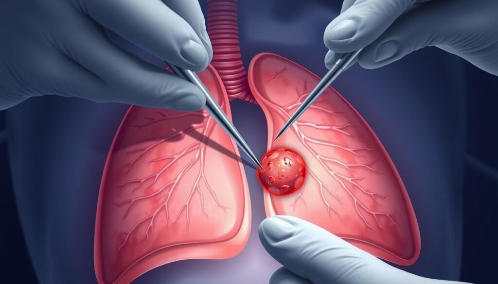
Needle biopsy techniques help get samples from the lung’s suspicious areas. These are often guided by CT scans or ultrasounds for accuracy. A thin needle is then used to take out cells for testing. This process is crucial for diagnosing lung cancer.
Bronchoscopy and Its Uses
Bronchoscopy lets doctors look directly at the airways to find tumors and get samples. It uses a flexible tube with a camera, inserted through the nose or mouth. This is especially useful for seeing central lesions and helps in diagnosing lung cancer accurately.

| Procedure | Description | Recommended Use |
|---|---|---|
| Needle Biopsy | Involves using a thin needle to collect tissue samples under imaging guidance. | Best for peripheral tumors. |
| Bronchoscopy | A tube with a camera is passed into the lungs to directly visualize airways. | Ideal for central tumors and biopsies. |
Clinical Decision Support in Lung Cancer Management
Clinical Decision Support Systems (CDSS) are key in lung cancer care, especially for small cell lung cancer (SCLC). The complexity of cancer cases makes oncology imaging very important. It helps make diagnosis and treatment plans better. This part talks about how clinical support uses imaging results. These results help choose the best treatment options.
The Role of Oncology Imaging
Oncology imaging offers deep insights into cancer. It helps tell different cancers apart, figure out the cancer stage, and create treatment plans. A study showed CDSS helps teams diagnose lung cancer right and match imaging results with clinic choices. Oncology imaging lets doctors double-check tests. This makes treatment plans more accurate.
How Imaging Guides Treatment Approaches
Choosing treatments for lung cancer depends a lot on imaging. A good CDSS points out when a treatment might not work for someone. It makes picking a personal treatment better. A CDSS gives a full look at a patient’s tests. This helps doctors talk over all important info. Knowing what imaging results mean leads to better decisions for lung cancer treatments. This hopes to make patient outcomes better. Learn more about how CDSS helps with managing symptoms here.
Understanding Pulmonary Nodules and Their Implications
Pulmonary nodules are small growths found in the lungs, often seen during chest X-rays or CT scans. They are detected about 150,000 times each year in the U.S. through X-rays. Their presence raises questions about lung cancer risk.
The use of chest CT scans has led to more lung nodules being found. From 2006 to 2012, detection rates increased from 3.9 to 6.6 per 1000 people. Yet, this increase hasn’t matched up with more lung cancer cases. This suggests most nodules are not cancerous. They are usually caused by past infections, scar tissue, or benign reasons.
Knowing what pulmonary nodules look like is important. Doctors use scans to check if a nodule grows over time. If it grows, they may use a PET scan for more details.

To find out if a nodule is cancerous, biopsy methods are used. The approach depends on where the nodule is located. If cancer is suspected, removing the nodule surgically might be the only option. Testing after the biopsy confirms the diagnosis. If cancer is found, more tests will outline the treatment needed.
If an infection is found, specialists may need to step in for treatment. Nodules must be watched over years with CT scans. This ensures any changes are noticed quickly. Understanding these steps helps health experts deal with pulmonary nodules and their possible link to lung cancer.
Staging and Treatment Options for SCLC
Small Cell Lung Cancer (SCLC) is mainly divided into two types: limited and extensive disease. The stage of the cancer greatly influences treatment options. Understanding the stage helps in predicting recovery and the overall outlook.
Limited vs. Extensive Disease
Limited disease means the cancer is only in one side of the chest. This allows for stronger treatment methods. On the other hand, 70-80% of people with SCLC are found to have extensive disease. This means the cancer has spread far in the body. Those with extensive-stage SCLC have a survival rate of just 2% after five years.
Surgery isn’t usually possible for those at this late stage. This fact underscores the importance of catching the disease early.
Importance of Staging in Treatment Decisions
The process of staging SCLC is vital for picking the right treatment. For those at the limited stage, doctors often use chemo combined with radiation. This method often works better than doing treatments one after another. Using chemo and radiation together is especially effective.
For extensive disease, treatment starts with chemo. Sometimes, doctors also use immunotherapy. To avoid brain tumors, they might use radiation on the head for those in the limited stage responding well to treatment.
| Staging | Treatment Options | Prognosis |
|---|---|---|
| Limited Disease | Chemotherapy + Radiation, Surgery (in rare cases) | Better outcomes, higher survival rates |
| Extensive Disease | Chemotherapy ± Immunotherapy | Poor prognosis, 5-year survival ~2% |
As medicine advances, discussing treatment options with doctors becomes crucial. Being informed about early detection and symptoms improves survival chances. For more info, read this critical resource.
Advancements in Thoracic Radiology
Recently, thoracic radiology has made big strides. This is due to new imaging tech that helps spot lung cancer better. Doctors can now make more precise guesses about health, thanks to these tools. This means they can catch the disease early and treat it faster.
New Imaging Technologies
New tools in lung imaging show major improvements in this field. For instance, a key breakthrough is the ultra-low-dose (ULD) CT scan. It cuts down on radiation, dropping it below 1 milliSievert. This reduction means folks are exposed to about 90% less radiation than before. The dose now is only around 0.15-0.2 mSv.
Studies on ULD in chest CTs focus on keeping pictures clear while spotting lung nodules well. Methods like tin filtration and special software show this effort. They also lower radiation by tweaking the CT scan settings. This includes changing the power settings and using smart algorithms.
Impact on Early Detection Methods
Dual-energy CT scans have become crucial, especially in improving image details. They help see blood flow in the lungs better. This is key in treating lung cancer better, knowing that 2.2 million people were diagnosed in 2020 alone.
Lung cancer is the top cancer in men and third in women worldwide. It was also the main cancer killer in 2020. These stats show why it’s vital to find and treat lung cancer early.

Conclusion
It’s vital to understand the role of x-rays in spotting small cell lung cancer (SCLC) early. This type of cancer makes up about 15% of all lung cancers. By using advanced x-ray techniques, we can find SCLC early, which greatly improves treatment and helps patients live longer. New ways of taking pictures of the lungs help find cancer sooner, leading to quicker treatment.
Every year, around 250,000 people worldwide are diagnosed with small cell lung cancer. This high number shows how important it is to keep looking for better ways to detect it early. Finding cancer early can mean a better chance of surviving. That’s why there’s a big push to improve how we take pictures of the lungs. Bringing these new methods into patient care is key in fighting lung cancer effectively.
Researchers are constantly trying to find better ways to look at the lungs and treat SCLC. There’s hope that these improvements will make life better for people with this disease. By focusing on better x-ray and imaging techniques, we’re paving the way for a future where we can beat lung cancer. This work gives hope to patients and the doctors treating them.