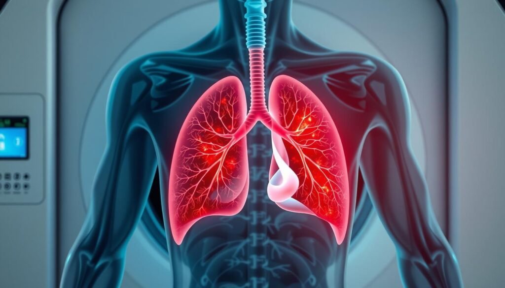
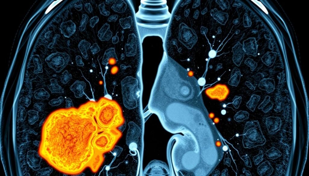
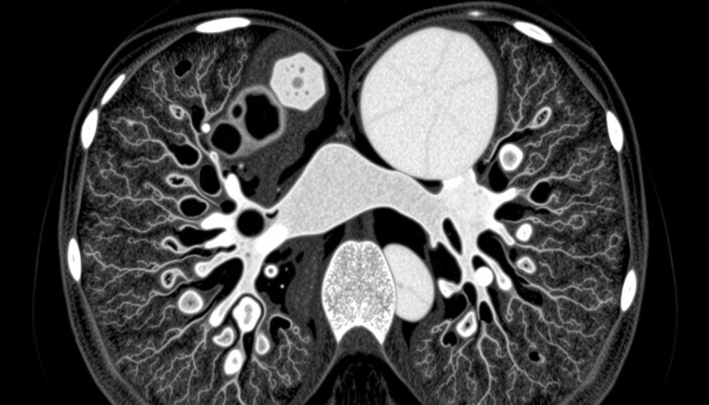
Did you know small cell lung cancer (SCLC) makes up about 13-15% of all lung cancer cases? This fact highlights how important SCLC is. It also shows how crucial accurate imaging like CT scans is for diagnosis and treatment. SCLC is a very aggressive lung cancer, often found when it’s already widespread. CT scans are key for doctors to assess this type of cancer. They help make well-informed choices about how to manage and treat patients.
Key Takeaways
- SCLC constitutes 13-15% of lung cancer cases, necessitating advanced imaging for effective management.
- CT scans play a vital role in lung cancer diagnosis by accurately visualizing lung structures.
- The median survival for untreated SCLC patients is alarmingly only 2-4 months.
- Imaging assists in identifying the extent of the disease, particularly limited vs. extensive SCLC.
- Approximately 70% of SCLC patients are diagnosed with extensive disease, emphasizing the need for prompt diagnosis.
Understanding Small Cell Lung Cancer
Small cell lung cancer (SCLC) is known for spreading quickly and starting early. It mainly affects those who smoke. This type results in 15-20% of all lung cancer cases. Understanding the spread of SCLC stresses the need for better awareness and treatments.
Overview of Small Cell Lung Cancer
About one out of three people found with SCLC have it in just one side of the chest. This is called limited-stage cancer. Most others have extensive disease, with cancer spreading far. The way doctors stage SCLC is different from other lung cancers. People with this cancer usually have a cough, feel short of breath, have chest pain, and lose weight. Spotting these signs early on is key.
Prevalence and Characteristics
Most people with lung cancer are diagnosed when the disease has spread widely. This makes treatment tough. Chemotherapy and radiation therapy are vital for all patients, though. Surgery is rare for those in the early stages of SCLC. Prompt action is crucial. For more details on SCLC and treatment methods, check this link.
The Role of Imaging in Lung Cancer Diagnosis
Imaging is very important in finding and treating lung cancer. Getting the right diagnosis is crucial. It helps doctors decide on the best treatment and affects patient outcomes. Many tools are used in imaging to find out if someone has lung cancer, what kind it is, and how far it has spread. This helps in choosing the right treatment.
Importance of Accurate Diagnosis
Knowing exactly what kind of lung cancer a patient has is key to treating it right. The five-year survival rate for lung cancer patients is only 15%. This shows why it’s so important to find the disease early and accurately. Most patients are already in an advanced stage when found, with fewer than 10% showing no symptoms at diagnosis. Better imaging means finding the cancer earlier. This could lead to better chances of surviving by starting treatment sooner.
Types of Lung Cancer Imaging Modalities
Doctors have many ways to look for lung cancer, each with its own benefits. The most common methods are:
- Chest X-rays: This is usually the first test but may not catch early-stage disease.
- CT Scans: They are key for finding out how far cancer has spread. They give detailed pictures of the lungs.
- MRI: Good for checking if cancer has spread to the brain or spine.
- PET Scans: They show the difference between cancerous and non-cancerous cells by looking at how they use energy.
Especially, CT scans are crucial in lung cancer diagnosis. They are better at spotting early-stage cancer compared to X-rays. CT scans can find small nodules in the lungs. Knowing the stage of cancer helps in predicting how well a patient might recover.
What is a CT Scan?
A CT scan, or computed tomography scan, makes detailed pictures inside the body using X-rays. It’s a non-invasive way to see organs and tissues. Doctors use it to get a clear view inside the body.
How CT Scans Work
CT scans work by taking many X-ray pictures from different angles. A computer then combines these pictures to make a detailed image. This method is great for spotting tumors and lesions. Using a special dye, or contrast, can make these images even clearer.
Benefits of CT Scans in Oncology
CT scans are vital in cancer care. They provide detailed info on tumors’ size and position. This is crucial for surgery planning and radiation therapy.
CT scans can find cancer early, which can save lives. They are better than standard X-rays for detecting cancers like lung cancer. This accuracy helps doctors monitor treatment and adjust it as needed.

| CT Scan Features | Description |
|---|---|
| Image Quality | High-resolution cross-sectional images of internal structures. |
| Versatility | Effective for diagnosing various cancers, including lung cancer. |
| Non-Invasive | No need for surgical procedures for imaging. |
| Contrast Usage | IV contrast enhances visibility of tumors and tissues. |
| Speed | Rapid imaging process, often completed within minutes. |
Small Cell Lung Cancer CT Scan
CT scans are crucial for diagnosing SCLC. They let doctors see unusual masses in the lungs. They also check for cancer spread to lymph nodes and nearby areas. An initial small cell lung cancer CT scan with contrast is standard. This makes the scan clearer.
How CT Scans are Used in Diagnosing SCLC
CT scans are key in spotting SCLC early. Each year, about 34,000 people in the U.S. find out they have SCLC. Sadly, most are already in the late stages. Early stages are rare, making up just 10.5% of cases. This situation underscores the need for better imaging to catch cancer early. Low-dose CT scans are especially hopeful, catching cancer early 76.5% of the time. Early detection helps doctors tailor treatment better. For more on imaging’s role, see this resource.
CT Scan and Staging of SCLC
Staging SCLC accurately is vital for choosing the right treatment quickly. CT scans are good at showing if the disease is limited or widespread. This helps doctors make important treatment choices. Plus, PET/CT scans bring even more precision, pointing out cancer spread to places like the liver and brain. With most SCLC found late, imaging’s role is clear. It guides oncologists in planning the best treatment path. For more on how diagnostics work, look at this guide.

CT Imaging Features of Small Cell Lung Cancer
Understanding CT images of Small Cell Lung Cancer (SCLC) is key to proper diagnosis and planning treatment. Imaging techniques help doctors see signs that show SCLC’s presence and how far it’s spread. This helps them fight this tough cancer better.
Common Findings on CT Scans
On CT scans, SCLC shows up in specific ways. Some familiar signs are:
- Centrally located masses: Often, SCLCs are big masses in the lung’s center, near important airways.
- Mediastinal involvement: These cancers tend to spread to nearby areas in the chest.
- Lymphadenopathy: Swollen lymph nodes, especially around the lungs and chest, signal the disease is advancing.
- Pleural effusions: Fluid in the lung area also suggests SCLC, together with other signs.
Classification of SCLC on Imaging
Categorizing SCLC on imaging is critical for choosing the best treatment approach. The focus is on where the tumor is and if it’s spread to the mediastinum. CT scans give doctors detailed info to plan the treatment that fits each patient’s situation.

Guidelines for Performing a CT Scan
Getting ready for a CT scan needs a few key steps for the best results and safety. Make sure you tell your healthcare provider if you have any allergies, especially to iodine-based contrasts. This step helps lower the risks from contrast media used in the scan.
Preparation for the CT Scan
To get a clear image from a CT scan, patients must follow certain rules. These prep steps are crucial:
- Notifying healthcare providers about allergies.
- Discussing recent medications and how they might affect the scan.
- Fasting for a few hours before the test if using contrast.
- Dressing comfortably and taking off metal objects to avoid image issues.
- Following any specific instructions from the healthcare team.
Contrast Use and Safety Considerations
Contrast media in CT scans make it easier to see tissues and spot problems. The American College of Radiology suggests IV contrast for better images in cancer checks. But, it’s important for patients to know about the safety of using contrast in CTs:
- Assessing renal function is vital for people with kidney issues.
- Monitoring for allergic reactions to the contrast is a must.
- Hydration before and after the scan helps avoid harm to the kidneys.
- Consulting radiologists if you’ve had a bad reaction to contrast before.
By following these CT scan prep tips and understanding contrast safety, patients help get precise diagnostic results.
Other Imaging Techniques
In the world of imaging techniques in oncology, MRI and PET/CT are key. They offer special insights that help assess lung cancer better. This support is vital for making decisions on treatment.
MRI and Its Role in Lung Cancer Evaluation
MRI is especially good at spotting brain metastases in lung cancer patients. It’s better than CT scans for telling benign from malignant lesions. MRI’s clear images of soft tissue are crucial in complex cases, even though it’s not the main way to look at lung cancer.
It becomes critical in some situations. It helps doctors be more accurate in their diagnoses and plan treatments.
Benefits of PET/CT Scans for SCLC
PET/CT scans bring a powerful method to see the metabolic activity of tumors in SCLC. These scans are key for the first diagnosis and staging. They offer details that other images can’t.
This combination gives a complete view. It lets doctors see how well treatments are working and monitor the disease’s path. Using PET/CT can catch disease spread early, vital for choosing the best treatments.
Treatment Planning Based on Imaging Results
Imaging is key when planning treatment for small cell lung cancer (SCLC). It helps find the tumor’s location and spread. Techniques like CT and PET scans allow doctors to create a customized treatment plan.
The staging from imaging helps decide the right therapy. This could be chemotherapy or radiation, among other treatments. Accurate treatment plans can improve outcomes, especially in early-stage disease that might be cured.
How Imaging Guides Treatment Options
Imaging results are crucial in choosing treatments for SCLC patients. Those with early disease could see better survival rates with chemo and radiation together. On the other hand, patients with advanced disease could improve their quality of life with specific treatments based on imaging.
This highlights how critical imaging is in care planning. It helps personalize treatments to offer the best care.
Integration of Imaging in Multidisciplinary Care
Managing SCLC well means embracing teamwork. Experts in radiology, oncology, and surgery must talk to each other well. They need to ensure that imaging results help guide the overall plan.
This cooperation helps doctors work together better. It can lead to improved care for patients. Focusing on teamwork in cancer care can help make better decisions and boost outcomes.
For more information on how imaging affects SCLC treatment plans, click here.