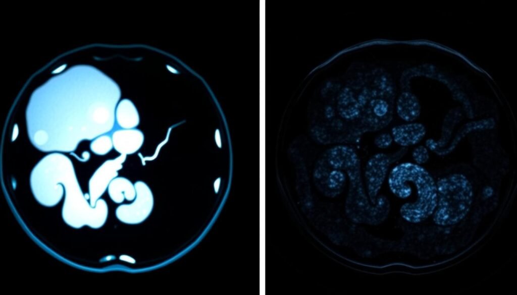
Since 1998, Medicare has covered many PET scan uses. This shows how important they are in finding cancer. It’s key for both patients and doctors to understand these scans well. This understanding helps in making the right choices for treatment because of how common cancer is becoming.
People often meet PET scans during health check-ups. So, it’s critical to know how these scans work and what their results mean. This part shares basics on reading PET scan outcomes. It especially focuses on what sets apart normal from cancerous scans.
Key Takeaways
- Understand the significance of PET scans in cancer detection.
- Differentiate between normal and cancerous PET scan results.
- Recognize the implications of accurate PET scan interpretations.
- Learn about the coverage history of PET scans in the United States.
- Appreciate the role of diagnostic accuracy in effective treatment management.
Understanding PET Scans
A PET scan stands for positron emission tomography. It’s key in today’s medical imaging. Learning what a PET scan is helps us understand its role in finding different health issues, especially cancers. Radioactive tracers are used in this process. They show the chemical activity in the body, giving doctors vital information.
What is a PET Scan?
A PET scan is a special method used in medicine that watches how the body reacts to a radioactive substance. This substance is usually a sugar-like material known as Fluorodeoxyglucose (FDG). After it’s injected, this tracer gathers in body tissues, highlighting active areas. Since problems like tumors absorb more tracer, they show up brighter in the images.
How Does a PET Scan Work?
The PET scan starts once the patient gets the radioactive tracer. The process takes between 25 to 50 minutes. During this time, the patient must stay still while the machine takes pictures from different sides. These pictures show changes in cells and their activity. This helps doctors not only find tumors but also learn how active they are and if they might be cancerous. By using PET scans with CT scans, doctors get a more complete view, improving diagnosis accuracy.
| Aspect | Details |
|---|---|
| Procedure Type | Nuclear medicine imaging technique |
| Radioactive Tracer | Fluorodeoxyglucose (FDG) |
| Typical Scan Duration | 25 to 50 minutes |
| Usage | Diagnosis and treatment monitoring of cancers, heart, and brain issues |
| Imaging Angles | Axial, sagittal, and coronal |
The Importance of PET Scans in Cancer Detection
PET scans are vital for spotting cancer early. This early detection is key in fighting cancer. They check cell activity that normal scans can’t. An unusual increase in this activity might mean cancer, leading to quick action.
Assessing Metabolic Activity
PET scans are good at seeing how active cells are. Cancer cells are usually busier than healthy ones. A special dye, called fluorodeoxyglucose (FDG), is used. It lights up active areas, showing where tumors might be. This can catch cancer sooner than CT or MRI scans, which look at the body’s structure.
Combining PET with Other Imaging Techniques
Mixing PET scans with other methods like PET-CT or PET-MRI improves accuracy. PET scans show cell activity. CT and MRI scans provide details about the body’s structure. With both, doctors get a full picture to create the best treatment plans. This approach is used in leading cancer centers. It helps in measuring tumors, checking for spread, and tracking how well treatments are working.
To learn more about detecting cancer, and the benefits of PET scans, many resources exist.
Normal vs Cancerous PET Scan Results
It’s vital to know the difference between normal and cancerous PET scan results. This part talks about what normal results look like. It also covers how to spot signs that might mean cancer.
What Constitutes Normal Results?
A normal PET scan shows the radiotracer spread out right in areas like the liver, brain, and spleen. There are no bad growths. This means the tissue is working fine, showing no cancer signs.
The scan won’t show weird radiotracer uptake in these spots. It proves cancer hasn’t moved from its original spot.
How to Identify Cancerous Results
Cancerous results on a PET scan have clear abnormalities, like bright spots. These spots mean there’s a lot of metabolic activity, which could be tumors. This could suggest cancer has spread or there’s a problem with how an organ works or infections.
Getting these differences right when looking at scans is key for a correct diagnosis.

| Type of Result | Description | Significance |
|---|---|---|
| Normal Results | No abnormal uptake in organs | Indicates no cancer spread |
| Cancerous Results | Bright spots indicating high FDG uptake | Potential malignancy or spread of cancer |
| False Positives | Abnormal results due to inflammation | Can mimic cancerous growths |
| Normal Variation | Physiological uptake patterns in some organs | Not indicative of disease |
Interpreting PET Scan Reports
Understanding PET scan reports might seem hard due to many medical terms and acronyms. It’s important for patients to know these terms to understand their results. One key term is the Standardized Uptake Value (SUV). This measures how much radiotracer, usually FDG, is in the tissues checked. A high SUV might mean there’s a lot of metabolic activity, which could signal cancer, inflammation, or infection.
Key Terms and Acronyms
Knowing these acronyms helps you talk better with your doctor. Here are some important terms about PET scan reports:
- FDG: Fluorodeoxyglucose, a radioactive glucose compound used in PET scans.
- SUV: Standardized Uptake Value, a measure of FDG concentration in the tissues.
- PET: Positron Emission Tomography, the imaging technique itself.
- CT: Computed Tomography, often combined with PET for more detailed imaging.
- MRI: Magnetic Resonance Imaging, another technique used in conjunction with PET scans.
Reading the Standardized Uptake Value (SUV)
The SUV tells us a lot about the tissue’s metabolic processes. High values mean more metabolic activity which could lead to various conditions, including cancer. It’s key to understand what the SUV means in your report. Look at the table below to see what different SUV values mean:
| SUV Range | Interpretation |
|---|---|
| 0.0 – 2.5 | Likely benign; normal metabolic activity. |
| 2.6 – 5.0 | Possible inflammation; requires further investigation. |
| 5.1 – 10.0 | Increased metabolic activity; consider the possibility of malignancy. |
| 10.1 and above | High probability of cancerous growth or aggressive disease. |
By understanding these terms and what SUV means, you can talk about your PET scan reports with your doctor more confidently.

Understanding FDG Uptake in Detail
Fluorodeoxyglucose, or FDG, is crucial in PET scans as a radiotracer. This substance, similar to glucose, shows how cells use sugar. It helps doctors understand how our body’s cells work. Knowing about FDG uptake gives clues about cellular functions. This is key in finding and figuring out diseases.
What is FDG and Its Role?
FDG is vital for imaging and finding diseases, like cancer. It acts as a radiotracer, showcasing tissue metabolism. High FDG levels in certain spots may mean faster metabolism, pointing to cancer. This trait makes FDG PET scans very useful. They help in identifying cancers in lungs, breasts, and colon, and in tracking how well treatments are working.
Normal, Mild, and Increased FDG Uptake
FDG uptake is broken down into levels, each showing different metabolic activities:
| FDG Uptake Level | Description | Implications |
|---|---|---|
| Normal | No significant uptake in metabolically inactive tissues | Shows tissues are working fine; like in the spleen and liver |
| Mild (SUV | Lower levels of FDG uptake | Often normal for less active tissues, but context is needed |
| Increased (SUV > 5) | Higher levels of FDG uptake | Suggests active metabolic processes, needs more checking |
It’s important to understand how FDG uptake relates to metabolic health. High FDG does not always mean cancer. Things like inflammation and infection can also raise levels. On the other hand, no uptake might show normal tissue working or possible issues.

Common Misinterpretations of PET Scans
Interpreting PET scans can be tricky, leading to important decisions for patient care. Knowing how to tell if lesions are harmless or harmful is key to correct diagnosis. Also, being aware of common issues like false positives and negatives helps everyone.
Differentiating Benign vs Malignant Lesions
One big issue with PET scans is telling apart benign from malignant lesions. Benign conditions can look active, like they’re taking in a lot of the scan’s tracer. This happens because of infections, inflammation, or changes after surgery. Such cases may show a scan suggesting cancer when there isn’t any. This can worry patients unnecessarily and lead to wrong treatment choices.
Understanding False Positives and False Negatives
False positives make healthy conditions seem cancerous. Things like blood sugar levels and infections can mess with scan results. This makes healthy tissue seem like cancer. False negatives, on the other hand, miss some cancers because they don’t show much activity. For example, low-grade lymphomas or small tumors might not be seen on a PET scan. This can delay treatment. Research says it’s important to check carefully and do more tests if needed to avoid wrong interpretations. Using different tracers, like PSMA, has shown to detect certain cancers better (source).
| Aspect | Benign Lesions | Malignant Lesions |
|---|---|---|
| Characteristics | Non-cancerous growths | Cancerous growths |
| Common Causes of False Positives | Infections, inflammation, post-surgical effects | N/A |
| Common Causes of False Negatives | N/A | Superficial tumors, low-activity tumors |
| Detection Techniques | Conventional imaging, follow-up | Advanced tracers like PSMA, combination testing |
With better understanding, patients can discuss their PET scan results more knowledgeably with their doctors.
The Role of Radiologists in PET Scan Interpretation
Radiologists are key in interpreting PET scan results. They provide vital insights for diagnostic accuracy. Their skill lies in reading PET images combined with knowing the patient’s history and symptoms. This ensures a full review. They understand the context of each scan, which is crucial. Because the same scan results can mean different things for different patients.
Diagnostic Accuracy and Clinical Context
In reading scans, radiologists with oncology expertise boost the trust in findings. Research shows that expert radiologists are more accurate than general doctors. Their in-depth knowledge aids in properly staging cancer and monitoring treatment. They notice details that others may miss, leading to better and more reliable diagnostics.
When Are Additional Tests Necessary?
At times, more tests are needed to verify a PET scan or clear up confusion. This might happen if results are unclear or symptoms and scans don’t match. Getting a second opinion from a cancer imaging specialist can help. Radiologists have different skills and experiences. An extra review can ensure more precise patient care.
Preparing for Your PET Scan
Getting ready for a PET scan is crucial. Knowing what steps to take makes the process better and more effective.
How to Get Ready
It’s important to get ready for a PET scan properly. Avoid eating 4 to 6 hours before your scan. Only drink water during this time. Also, don’t do heavy exercise for six hours before your scan to keep your results clear.
Tell the clinic about any medicines or allergies you have. Also, let them know if you’re pregnant. Remember to take off any metal objects, as they can mess up the scan.
What to Expect During the Procedure
They will inject a special sugar into your body for the PET scan. This sugar helps them see your body better. You’ll need to wait about an hour for your body to absorb it. The scan itself lasts about 30 to 45 minutes.
After the scan, you can do your usual activities. But make sure to drink lots of water to get the sugar out of your system. You’ll get your scan results in one or two weeks. If it’s taking longer, call your doctor. For more tips, check out this guide on preparing for your PET.
Conclusion
PET scans are key in finding cancer. They offer deep insights, unlike traditional imaging. By spotting metabolic changes in tumors, they help catch cancer early. This early detection plays a big role in choosing treatments. Studies show that PET scans were helpful in 67% of cases where patients had normal CEA levels but might have had a tumor come back. This proves their worth in different medical situations.
Understanding PET scan results is vital. It helps doctors manage a patient’s care better. With a 76.9% accuracy rate in spotting tumor return, doctors and radiologists can make smarter choices. After PET scans, doctors changed their plans for 38.0% of patients. This highlights how important these scans are in deciding on treatments.
While PET scans are useful, they’re complicated and need a team effort. For more on PET technology and its use in cancer care, click here. Working together helps patients and doctors deal with cancer more effectively.