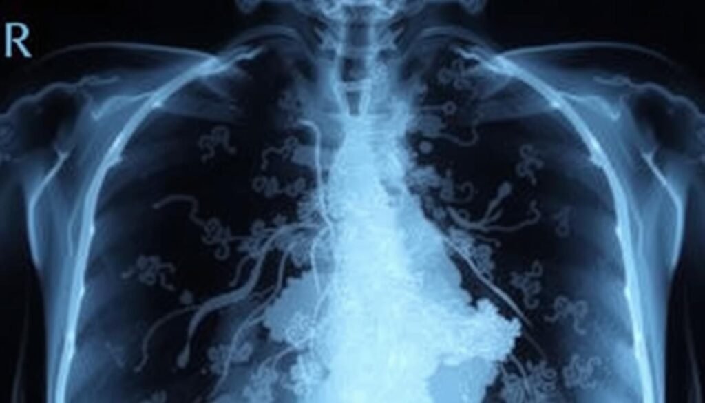
Staggeringly, an estimated 238,340 individuals in the United States will get lung cancer by 2023’s end. This shows how crucial effective diagnostic tools are, especially early on. Chest X-rays play a key role as starting tests to spot abnormalities in the lungs.
Spotting lung cancer early on X-ray can greatly improve treatment chances. X-rays let us see inside the lungs to find unusual masses or tissue changes. But, lung cancer often doesn’t show symptoms until it’s late. That’s why regular screenings are critical, especially for those 50 to 80 who smoked or still smoke.
We’re now going to look deeper into how X-rays help find lung cancer. We’ll talk about how they work, their limits, and why we need more tests to be sure.
Key Takeaways
- Chest X-rays are commonly used as initial tests to diagnose lung cancer.
- Yearly lung cancer screening is recommended for high-risk groups.
- Identifying lung cancer on X-rays can lead to earlier treatment options.
- Only about 10-15% of lung cancers are Small Cell Lung Cancer (SCLC).
- Soft tissues appear darker on X-rays, complicating the identification of some tumors.
Introduction to Lung Cancer Diagnosis
Diagnosing lung cancer starts when you notice certain lung cancer symptoms. These can include a lingering cough, shortness of breath, chest pain, and losing weight without trying. If you have these symptoms, it’s important to see a doctor. Share your health history and any risk factors like smoking or being around harmful substances. This helps in figuring out if it’s lung cancer.
The first step might be getting a chest X-ray to check for problems in the lungs. Sometimes, though, these X-rays don’t tell us everything. They can’t always show if an issue is cancer or something else. If the X-ray looks odd, the doctor might ask for more tests. This could mean blood tests or checking how well your lungs work.
Often, after an X-ray, you might need a CT scan. This gives a much clearer picture of your lungs. If needed, even more precise tests like PET-CT scans can spot cancer cells. This helps doctors decide the best way to treat it. If they’re still not sure, you might get a bronchoscopy or a biopsy. These tests take samples from your lungs for closer examination. They are key in making a final lung cancer diagnosis.
The Role of X-Rays in Lung Cancer Detection
X-rays are a critical first step in lung cancer imaging. They use radiation to create lung images. This method looks for differences in tissue density to find problems. However, the role of x-rays in cancer detection has limits. They might miss small or widespread cancers if used alone.
About 90% of lung cancer misdiagnoses involve chest x-rays. In contrast, only 10% were with CT scans and advanced tools. A study found reviewing radiographs without rushing aids in spotting lesions better. Also, comparing radiographs side-by-side helps find hidden lesions sooner. This shows why we need more than one imaging method.
Chest x-rays are easy to use and give off little radiation. They’re commonly used for checking lung issues, even though they’re not perfect at finding cancer. When x-rays show abnormal spots as white-grey, further tests like CT scans and bronchoscopy are needed. X-rays start the diagnosis process, leading to more accurate methods.

What Does Lung Cancer Look Like on an X-Ray
Imaging techniques are vital for diagnosing lung cancer. They help us see lung cancer on an x-ray, influencing treatment options. When checking X-rays for lung tumors, specific features stand out.
Understanding Radiographic Appearance
Normally, lungs look dark gray on an X-ray because they’re full of air. Lung tumors, however, show up as lighter spots. This difference helps experts find areas that might have cancer. Roughly 80% of lung cancers are called non-small cell lung cancer (NSCLC). This group includes types like adenocarcinoma and squamous cell carcinoma, which have unique shapes. Small cell lung cancer (SCLC) makes up about 13% of cases. Its tumors can look different because of how the cells are structured.
Typical Features of Lung Tumors
Lung tumors differ in appearance based on their kind. For instance, adenocarcinoma often appears as uneven, nodular shadows. In contrast, squamous cell carcinoma might look more like flat irregularities. These are usually in the lung’s outer parts. SCLC tumors are seen as small oval shapes and are sometimes called oat cell cancer.

Common X-Ray Findings Associated with Lung Cancer
Lung cancer comes with many risk factors and symptoms. Spotting it early is critical. Radiologists look for specific lung cancer x-ray findings to find cancer early. Knowing about these findings can lead to early treatment and better chances of survival. This section will cover lung cancers that can be seen on X-rays and their signs.
Types of Lung Cancer Visible on X-Rays
There are two main lung cancers seen in X-rays: Small Cell Lung Cancer (SCLC) and Non-Small Cell Lung Cancer (NSCLC). Let’s explore their differences:
- Small Cell Lung Cancer (SCLC): Makes up about 15-20% of lung cancer cases, mainly found in smokers.
- Non-Small Cell Lung Cancer (NSCLC): This kind is 80-85% of lung cancers. It includes types like:
- Squamous Cell Carcinoma: Found mostly in the center of the lungs, making up about 30% of cases.
- Adenocarcinoma: Usually seen in non-smokers, especially young women.
- Large Cell Carcinoma: Linked to smoking and notable for significant damage.
Characteristics of Abnormalities
Radiologists check X-rays for clear signs of lung cancer. Some key x-ray characteristics of lung cancer are:
- Lung Nodules: Small, round spots on the X-ray, often needing more tests.
- Lung Masses: Big, uneven shapes might mean the disease is far along or has spread.
- Cavitary Lesions: Though not as common, these suggest tumors that are dying or infections.
- Miliary Nodules: Symbolic of cancer spreading, they look like small, regular shadows.

Limitations of Chest X-Rays in Diagnosing Lung Cancer
Chest X-rays have big challenges when it comes to spotting lung cancer. Studies show their sensitivity is about 76.8% to 79.7%. This means many lung cancer cases might not be caught early on.
Overlapping conditions like pneumonia or tuberculosis can hide cancer signs. Small tumors can be missed behind bones or tissues. Roughly 20% of lung cancer cases are not found in chest X-ray screenings.
The quality of research on the effectiveness of X-rays is often poor. Of 21 studies looked at, most had a high risk of bias. Only a few were more reliable.
Doctors face tough odds with X-ray detection. A chest X-ray finds tumors around 20 mm big. But a CAT scan can spot tumors as small as 5 mm. This shows why advanced imaging is key for those at high risk.
There’s a real need for better training for healthcare workers on reading chest X-rays. Better training can help catch more cases early. Lung cancer causes many cancer deaths, so accurate diagnosis is crucial.
More research is needed to see which patients need more tests after a negative X-ray. Using things like low-dose CAT scans could help find more cases early. This could tackle the weaknesses of chest X-rays.
To learn more about these challenges, check out this literature here.
Alternative Imaging Techniques for Lung Cancer Diagnosis
Lung cancer diagnosis has come a long way, thanks to improved imaging techniques. Instead of just X-rays, doctors now have CT scans lung cancer diagnosis and PET scans lung cancer. These options offer clearer images and details.
CT scans produce detailed images of the lungs. This allows doctors to see abnormalities clearly. Compared to chest X-rays, CT scans are more accurate. They play a key role in understanding lung cancer stages.
Systems like the Brock model use CT scans. They look at nodule size and location to predict if a tumor is malignant.
PET scans use a special dye to spotlight cancer cells. They’re great for finding active tumors and checking for spread. When PET and CT scans are combined, the result is even more precise. This helps in accurately detecting lung cancer.
CT scans are particularly useful because they guide biopsies. This ensures doctors take samples from the right spots. Using CT-guided biopsies is effective and reduces risks. Imaging in lung cancer diagnosis is crucial. It supports early assessments and leads to thorough evaluations.
Want more information on diagnosing lung cancer? Check out different imaging techniques through this informative link.
Identifying Lung Cancer: Other Tests and Procedures
For lung cancer, doctors use imaging and biopsy methods to diagnose correctly. Chest X-rays are a first step but don’t show everything. CT scans give a much clearer picture of lung tumors.
CT and PET Scans Compared to X-Rays
CT scans are better than X-rays for finding lung tumors. They show detailed images of the lungs. PET scans are also crucial. They help figure out if the cancer has spread. This is important for choosing the right treatment. MRI scans are helpful, too. They are used to see if cancer reached critical areas like the brain or liver.
Biopsy Procedures for Definitive Diagnosis
When scans suggest lung cancer, more tests are needed to be sure. There are several biopsy methods. Fine needle aspiration (FNA) biopsies are used for small masses or lymph nodes. Core biopsies get larger samples and give more information. Thoracentesis removes fluid from around the lungs to check for cancer. Sputum cytology can help find certain lung cancers, like squamous cell carcinoma.
It’s vital to detect lung cancer early. Knowing the early signs can lead to quicker help from doctors. For more on early lung cancer signs, visit this link.
Monitoring Lung Cancer Treatment Progress with X-Rays
X-rays are key in tracking lung cancer treatment. They help doctors see how effective the treatment is. Doctors use them to observe tumor size changes and new lung growths during and after treatment. It’s vital to have x-ray check-ups regularly for patient care.
Knowing how a patient is recovering is crucial for planning further treatment. X-rays quickly show any changes in the tumor. This shows if the treatment works or if plans need changing. Keeping a regular check-up schedule helps catch any tumors that come back after the first treatment.
Quick action is often necessary for lung cancer treatment, making x-ray follow-ups essential. While experts debate how often patients should be imaged, it’s clear that close monitoring saves lives. Recent studies suggest new ways to improve how we monitor lung cancer treatment. For more details, check this source.
| Treatment Stage | 5-Year Survival Rate |
|---|---|
| Local Stage | 63% |
| Regional Stage | 35% |
| Distant Stage | 8% |
Conclusion
Early detection is key in fighting lung cancer. Chest X-rays are vital for spotting issues early on. They let doctors see if there’s anything wrong in the lungs or nearby areas. But, these lung cancer x-ray findings need more tests to make sure they’re right.
People should watch out for signs like constant coughing or losing weight without trying. These could be early warning signals of lung issues. It’s crucial to talk to doctors when these symptoms show up. Doing this early can really help the chances of getting better. Plus, knowing the warning signs of lung cancer helps people get help faster.
Making sure we catch lung cancer early is our main aim. This needs everyone to keep an eye out for symptoms and to have open talks with doctors. With early spotting and good communication, we can fight lung cancer together and aim for the best health results.