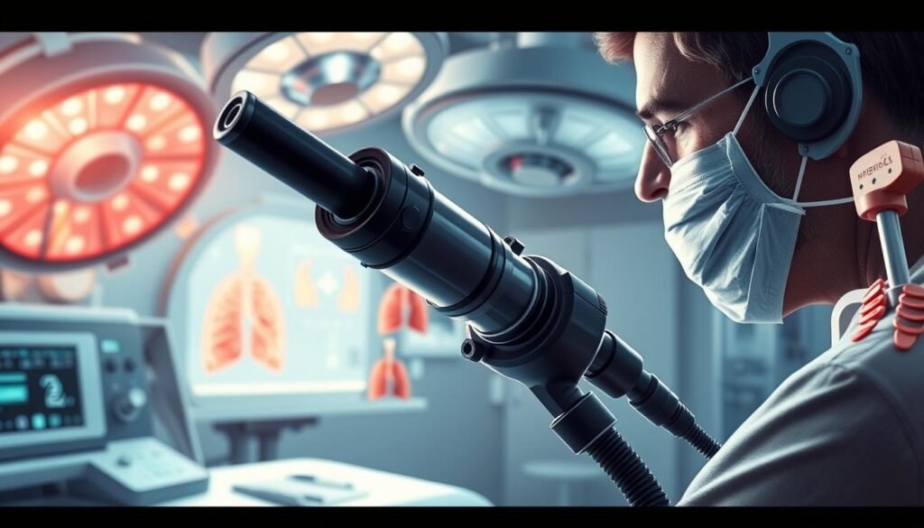
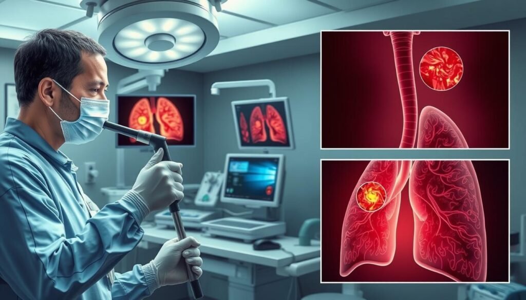
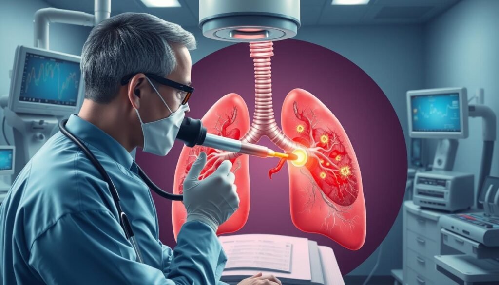
Did you know over 228,000 new lung cancer cases are expected this year in the US? This highlights the need for effective diagnostic tools, like bronchoscopy. Through a thin, flexible tube with a camera, doctors can explore the lungs. This method offers a close view into lung conditions, including cancer.
Bronchoscopy has grown to be less invasive, enhancing both reach and diagnostic precision. It’s crucial for catching lung cancer early, leading to hopeful paths for patients through timely care and tailored treatments. Bronchoscopy’s data is vital, helping doctors fight lung cancer with better strategies. For more on bronchoscopy, see the Mayo Clinic’s guide.
Key Takeaways
- Bronchoscopy is vital in the diagnosis and treatment of lung cancer.
- The procedure typically takes about 30-45 minutes to complete.
- Results from biopsies collected during bronchoscopy can take up to a week for final reports.
- Conscious sedation allows patients to breathe independently during the procedure.
- Some common side effects post-procedure include a sore throat, manageable with cough drops.
- Advanced techniques like autofluorescence bronchoscopy are being investigated for better diagnostic sensitivity.
- Monitoring equipment continuously tracks vital signs throughout the bronchoscopy procedure.
Introduction to Bronchoscopy
Bronchoscopy is key in studying lung diseases, offering a clear view inside the airways and lungs. It uses a flexible tool called a bronchoscope, which has a light and camera. Through the mouth or nose, this tool lets doctors see the respiratory path clearly, for detailed checks.
Doctors often use bronchoscopy to look into ongoing coughs, odd chest X-ray results, or possible lung infections. The technology, introduced in the late ’60s, changed how lung problems are diagnosed. Thanks to flexible bronchoscopes, these checks are less intrusive.
A bronchoscopy might last from 15 to 60 minutes, based on the examination’s depth. After, patients usually rest for about 45 minutes to check on vital signs and anesthesia effects. Most of these exams are outpatient, meaning they can go home the same day. This tool’s ability to extract crucial lung information is priceless in lung health.
Understanding the Bronchoscopy Procedure
The bronchoscopy procedure is key for diagnosing lung problems and offering treatments. It’s usually done in one visit, taking 30 to 60 minutes. Patients get sedation and local anesthesia to be comfortable. They need several hours to recover afterward.
A flexible bronchoscope lets doctors see the airways as it happens. This helps them get tissue and fluid samples for tests. These tests help find infections, tumors, or other lung issues.
The procedure can also treat certain lung problems. Doctors can clear blockages, drain fluid, or put in stents to keep airways open. Though rare, some might have mild bleeding, cough, or infection. It’s important to watch for serious symptoms like chest pain or trouble breathing afterward.
After the procedure, patients might have a sore throat or sound hoarse for a little while. Biopsy results are ready in a few days. Because of the sedation, patients should not drive home. They’ll need a ride instead.

How Bronchoscopy is Used to Diagnose Lung Cancer
Bronchoscopy is key for diagnosing lung cancer. It lets doctors see the airways and get important tissue samples. This helps find tumors, know where they are, and check for blockages in the lungs. Bronchoscopy is crucial for accurate lung cancer diagnoses.
The Role of Flexible Bronchoscopy
Flexible bronchoscopy stands out because it reaches the lungs’ farthest parts. Doctors can easily check harder-to-reach spots with this tool. They can also take samples of tissue during the procedure. This is key for confirming if the tissue is cancerous.
These samples help understand the tumor’s nature. They help create treatment plans that fit the patient’s needs.
Types of Bronchoscopes Used
Doctors use different kinds of bronchoscopes, like flexible and rigid ones. The flexible ones are preferred for their ease of use in the lung’s twists and turns. But rigid bronchoscopes are better for dealing with big blockages in the airways.
Each type helps doctors get a better look at lung health. This makes sure lung issues are thoroughly examined.

Indications for Bronchoscopy in Lung Cancer
Bronchoscopy is key for checking suspected lung cancer. It’s advised for patients with symptoms like persistent coughing, unexpected weight loss, or coughing up blood. If imaging tests spot issues, bronchoscopy offers deep insights. It helps figure out why these symptoms are happening.
Common Symptoms Leading to Bronchoscopy
Several warning signs suggest the need for a bronchoscopy in lung cancer:
- Abnormalities detected in lung imaging tests
- Biopsy of lymph nodes near the lungs
- Coughing up blood
- Shortness of breath
- Low oxygen levels
- Prolonged unexplained cough
- Unresolving lung infections
- Inhalation of toxic substances
For those with Chronic Obstructive Pulmonary Disease (COPD), it’s vital to recognize these symptoms. There’s a higher lung cancer risk. Knowing the common signs of COPD and lung cancer can help with early diagnosis and treatment. More details are available here.
Biopsy and Tissue Sampling
Bronchoscopy mainly collects tissue samples. During the procedure, different techniques help get these samples, like:
- Brushings
- Biopsy forceps
- Transbronchial needle aspiration (TBNA)
The samples are then checked, giving vital info about lung issues. This is crucial for diagnosing and deciding the right treatment in lung cancer.

To learn more about flexible bronchoscopy, check out the guidelines here. Bronchoscopy’s ability to clarify unclear imaging findings and directly sample tissue makes it essential in lung cancer care.
Advanced Bronchoscopic Techniques
Recent progress in bronchoscopic techniques has changed how lung cancer is diagnosed and managed. These new tools help doctors examine and treat lung problems better. The most noteworthy are endobronchial ultrasound (EBUS) and autofluorescence imaging.
Endobronchial Ultrasound (EBUS)
EBUS is a cutting-edge technique that lets doctors check the lymph nodes and lung lesions better. It uses ultrasound with a bronchoscope for live images, increasing diagnostic accuracy. Thanks to EBUS, the procedure called endobronchial ultrasound-guided transbronchial needle aspiration (EBUS-TBNA) has become highly successful. It shows about 90% success in lung cancer staging, almost as good as the older method, mediastinoscopy.
| Technique | Sensitivity | Agreement with Mediastinoscopy |
|---|---|---|
| EBUS-TBNA | 90% | 91% |
| Video Mediastinoscopy | 89% | N/A |
Autofluorescence and Narrow Band Imaging
Autofluorescence and narrow band imaging are great for finding early-stage lung cancers and precancerous lesions. They make it easier for doctors to see problems during bronchoscopic procedures. These methods show changes that regular methods could miss. Autofluorescence bronchoscopy, used with white light, improves the ability to detect lung cancer.
Risks and Complications of Bronchoscopy
Patients getting a bronchoscopy should know its risks and complications. Even though it’s usually safe, knowing what to expect helps. This knowledge can make the procedure smoother and safer.
Understanding Procedure-Related Risks
Bleeding at the biopsy site and infections are common risks of a bronchoscopy. Some people might get a fever shortly after. Serious problems, like a collapsed lung or major bleeding, are rare but can happen.
People on blood thinners need to pause these meds before their procedure. This is vital because bleeding risks go up with such medicines. The death rate from bronchoscopies, whether flexible or rigid, is very low, under 0.1 percent.
During the process, some might see their blood pressure change or feel nauseous. Very few might have heart rhythm problems or trouble breathing. Watching patients closely during bronchoscopy helps catch any issues early on.
Post-Procedure Monitoring
After a bronchoscopy, patients stay under watch. This helps spot any immediate issues fast. Trouble breathing, a fast heart rate, or chest pain need quick action.
Recovery often comes with a sore throat for a few days. Patients are advised to rest and go easy. Watching blood oxygen levels is key for those who had extra oxygen during the process.
Both healthcare workers and patients benefit from understanding bronchoscopy risks and complications. This knowledge leads to safer, more effective care.
Recovery and Aftercare Following Bronchoscopy
Recovery after a bronchoscopy is key for good healing. Patients often stay in a recovery area to be watched. They have to wait until they are fully alert before going home. The throat and mouth might feel numb for hours. This means eating and drinking should wait until the feeling comes back.
It’s common to feel a bit uncomfortable. You might have a sore throat or cough a little. Usually, this discomfort goes away on its own. To help with recovery, patient care includes following eating advice. You should try soft foods and drink more, as long as it feels okay.
After the procedure, it’s important to watch out for certain symptoms. These include:
- Excessive bleeding
- Difficulty breathing
- Persistent fever over 100°F
- Coughing up lots of blood
- Feeling lightheaded or dizzy
To reduce the risk of bleeding, don’t take aspirin or ibuprofen for the first day. Resting for the day helps recovery, especially if you were sedated. You shouldn’t drive, use machines, or sign papers during this time.
Two hours after, patients can start having sips of water. They can eat and drink normally if they can swallow without trouble. Feeling tired for a day or two, having a dry mouth, sore throat, or hoarseness are common. You might see a little blood in your saliva, especially if they did a biopsy. This is usually okay. It’s best to not smoke for at least a day post-procedure.
Following these tips helps ensure a safe recovery from a bronchoscopy. It’s very important to go to follow-up appointments for the best health results. For more details on aftercare, you can read these detailed instructions.
Conclusion
Bronchoscopy is key for finding lung cancer. It gives doctors a clear view and lets them get tissue samples. This is very important for deciding what the cancer is like and how far it has spread.
This tool’s importance is huge. It shows images in real time and helps get samples, which are very important for correct diagnosis. The advancements in this procedure make it safer and more effective. This leads to better health results for patients with breathing issues.
It’s also crucial to keep an eye on patients after their test, especially if their biopsy doesn’t show cancer. Problems like inflammation or blockages in the airways need close monitoring. Knowing these details helps doctors plan the best treatment for each patient. This approach aims for a hopeful outlook in treating lung cancer.
Using new methods along with traditional bronchoscopy helps doctors deal with lung cancer better. It speeds up finding the disease and starting treatment. This improves care quality in respiratory health. As treatments for lung cancer keep getting better, bronchoscopy is still a vital tool for helping patients achieve better health results.